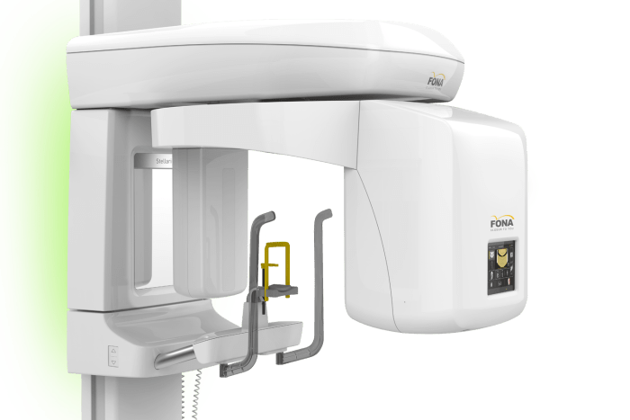
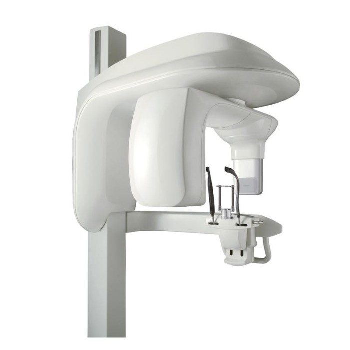
A grid used with extraoral imaging – Grids play a pivotal role in extraoral imaging, enhancing image quality by minimizing scattered radiation. Understanding the components, design, function, evaluation, and applications of grids is essential for optimizing imaging outcomes in various clinical scenarios.
Grids are composed of alternating layers of lead and radiolucent material, effectively absorbing scattered radiation while allowing primary radiation to pass through.
Grid Components

A grid used with extraoral imaging consists of a series of parallel lead strips separated by a radiolucent material. The lead strips absorb scattered radiation, while the radiolucent material allows the primary radiation to pass through.
Materials Used in Grid Construction
- Lead: Lead is a dense material with a high atomic number, which makes it effective at absorbing X-rays.
- Aluminum: Aluminum is a lightweight material with a lower atomic number than lead, which makes it less effective at absorbing X-rays. However, aluminum is more radiolucent than lead, which allows more primary radiation to pass through.
- Carbon fiber: Carbon fiber is a lightweight and strong material that is radiolucent. It is often used in the construction of grids for mobile imaging systems.
Grid Design
Grids are available in a variety of designs, including:
Parallel Grids
Parallel grids have lead strips that are arranged in parallel lines. This design is simple and effective, but it can cause some artifacts in the image.
Focused Grids, A grid used with extraoral imaging
Focused grids have lead strips that are curved to focus the primary radiation on the image receptor. This design reduces artifacts, but it is more complex and expensive to manufacture.
Cross-Hatched Grids
Cross-hatched grids have lead strips that are arranged in a cross-hatched pattern. This design reduces artifacts and scatter, but it is more complex and expensive to manufacture than parallel grids.
Grid Function: A Grid Used With Extraoral Imaging

A grid functions by absorbing scattered radiation. Scattered radiation is X-radiation that has been deflected from its original path by interactions with matter. This radiation can degrade the image quality by reducing the contrast and sharpness of the image.
Primary and Secondary Radiation
Primary radiation is X-radiation that has not been scattered. Secondary radiation is X-radiation that has been scattered. The grid absorbs the secondary radiation, but it allows the primary radiation to pass through.
Grid Evaluation

Grids are evaluated based on their ability to reduce scattered radiation. The grid ratio is a measure of the grid’s effectiveness in reducing scatter. The grid ratio is calculated by dividing the amount of scattered radiation with the grid in place by the amount of scattered radiation without the grid in place.
Factors Affecting Grid Evaluation
- Grid ratio: The grid ratio is the most important factor in grid evaluation. A higher grid ratio indicates that the grid is more effective at reducing scatter.
- Grid frequency: The grid frequency is the number of lead strips per centimeter. A higher grid frequency indicates that the grid is more effective at reducing scatter.
- Focal spot size: The focal spot size is the size of the X-ray beam at the source. A larger focal spot size results in more scattered radiation, which can reduce the grid’s effectiveness.
- Image receptor size: The image receptor size is the size of the X-ray film or digital sensor. A larger image receptor size results in more scattered radiation, which can reduce the grid’s effectiveness.
Grid Applications
Grids are used in a variety of extraoral imaging applications, including:
Dental Radiography
Grids are used in dental radiography to reduce scattered radiation from the patient’s teeth and jaws. This improves the contrast and sharpness of the image.
Chest Radiography
Grids are used in chest radiography to reduce scattered radiation from the patient’s lungs and heart. This improves the contrast and sharpness of the image.
Mammography
Grids are used in mammography to reduce scattered radiation from the patient’s breast. This improves the contrast and sharpness of the image, which can help to detect breast cancer at an early stage.
Grid Selection
The selection of a grid for extraoral imaging depends on a number of factors, including:
Imaging Application
The imaging application is the most important factor in grid selection. The type of grid that is used will depend on the specific imaging task.
Patient Size
The patient’s size is also a factor in grid selection. A larger patient will require a grid with a higher grid ratio and a higher grid frequency.
Focal Spot Size
The focal spot size is the size of the X-ray beam at the source. A larger focal spot size results in more scattered radiation, which can reduce the grid’s effectiveness.
Image Receptor Size
The image receptor size is the size of the X-ray film or digital sensor. A larger image receptor size results in more scattered radiation, which can reduce the grid’s effectiveness.
Common Queries
What is the primary function of a grid in extraoral imaging?
Grids reduce scattered radiation, which improves image contrast and reduces image noise.
What are the key factors to consider when selecting a grid for extraoral imaging?
Focal spot size, image receptor size, and the intended clinical application should be considered when selecting a grid.
How do grids affect image quality?
Grids improve image quality by reducing the amount of scattered radiation reaching the image receptor, resulting in improved contrast and reduced noise.