Correctly label the structures of the implanting blastocyst. – Correctly Labeling the Structures of the Implanting Blastocyst: Delving into the intricate complexities of the implanting blastocyst, this discourse unravels the fundamental principles that govern its structures, laying the groundwork for a comprehensive understanding of early embryonic development.
The initial stages of embryonic development, marked by the formation and implantation of the blastocyst, hold immense significance in shaping the trajectory of an organism’s life. This discourse embarks on an exploration of the intricate processes involved in blastocyst implantation, deciphering the roles of various structures and their contributions to the establishment of a viable pregnancy.
Embryonic Layers

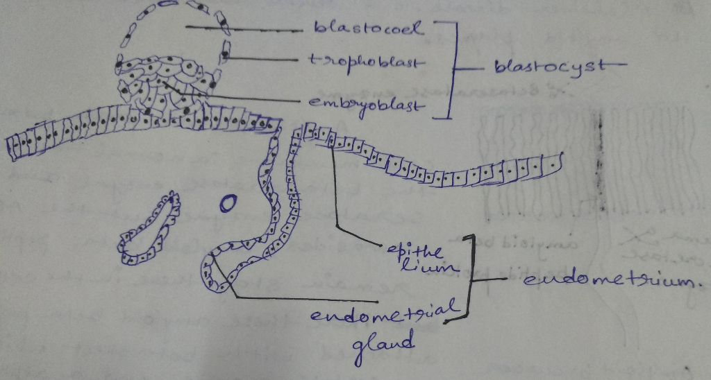
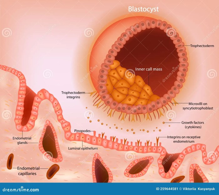
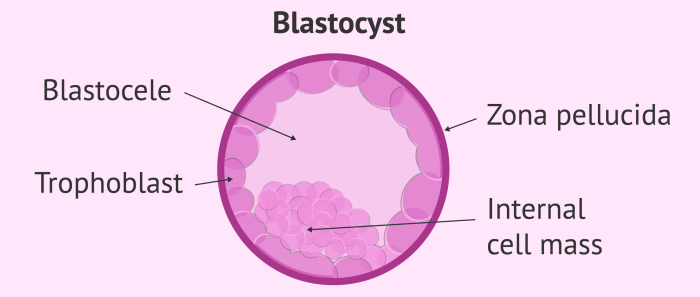
The blastocyst consists of two distinct layers: the inner cell mass (ICM) and the trophoblast. The ICM gives rise to the embryo proper, while the trophoblast forms the extraembryonic membranes that support and nourish the developing embryo.
Trophoblast
The trophoblast is the outer layer of the blastocyst. It consists of a single layer of flattened cells that are in direct contact with the uterine wall. The trophoblast plays a crucial role in implantation by secreting enzymes that dissolve the uterine lining and allow the blastocyst to embed itself within the uterine wall.
Inner Cell Mass
The ICM is located within the trophoblast and consists of a group of pluripotent cells that have the potential to differentiate into any type of cell in the body. As the blastocyst implants, the ICM differentiates into two layers: the epiblast and the hypoblast.
The epiblast gives rise to the embryo proper, while the hypoblast forms the yolk sac, which provides nutrients to the developing embryo.
Implantation Process: Correctly Label The Structures Of The Implanting Blastocyst.

Implantation is the process by which the blastocyst attaches to the uterine wall and begins to develop. The implantation process can be divided into three main stages:
Zona Pellucida Hatching
The first stage of implantation involves the hatching of the blastocyst from the zona pellucida, a protective layer that surrounds the blastocyst. The hatching process is mediated by enzymes secreted by the trophoblast.
Attachment to the Uterine Wall
Once the blastocyst has hatched from the zona pellucida, it attaches to the uterine wall. The attachment process is mediated by adhesion molecules expressed on the surface of the trophoblast and the uterine wall.
Invasion of the Uterine Wall
After the blastocyst has attached to the uterine wall, the trophoblast begins to invade the uterine wall. The invasion process is mediated by enzymes secreted by the trophoblast that dissolve the uterine lining. As the trophoblast invades the uterine wall, it forms a network of blood vessels that connect the developing embryo to the maternal circulation.
Formation of the Placenta

The placenta is a specialized organ that develops from the trophoblast and plays a crucial role in nutrient exchange and waste removal between the mother and the developing embryo. The placenta is formed by the fusion of the trophoblast with the uterine wall.
The placental villi, which are finger-like projections of the trophoblast, invade the uterine wall and establish a close connection with the maternal blood vessels. This close connection allows for the exchange of nutrients, oxygen, and waste products between the mother and the developing embryo.
Structure of the Placenta
The placenta consists of two main components: the maternal component and the fetal component. The maternal component consists of the uterine wall, while the fetal component consists of the chorionic villi. The chorionic villi are covered by a layer of trophoblast cells that are in direct contact with the maternal blood vessels.
Function of the Placenta, Correctly label the structures of the implanting blastocyst.
The placenta plays a crucial role in the development of the embryo. It provides nutrients and oxygen to the embryo and removes waste products. The placenta also produces hormones that are essential for the maintenance of pregnancy.
Embryonic Development

The early stages of embryonic development occur within the blastocyst. The first step in embryonic development is the formation of the embryonic disc. The embryonic disc is a flat, round structure that consists of two layers: the epiblast and the hypoblast.
The epiblast gives rise to the embryo proper, while the hypoblast forms the yolk sac.
Primitive Streak
The primitive streak is a thickened region of the embryonic disc that appears during gastrulation. Gastrulation is the process by which the three germ layers of the embryo (the ectoderm, mesoderm, and endoderm) are formed. The primitive streak gives rise to the mesoderm, which forms the muscles, bones, and connective tissues of the body.
Extraembryonic Membranes
The extraembryonic membranes are a group of membranes that surround the embryo and play a crucial role in protecting and nourishing the developing embryo. The extraembryonic membranes include the amnion, chorion, and yolk sac.
Amnion
The amnion is a thin, transparent membrane that surrounds the embryo and fills with fluid. The amniotic fluid provides a protective environment for the embryo and allows it to move freely.
Chorion
The chorion is a vascular membrane that surrounds the amnion and the embryo. The chorion is responsible for the exchange of nutrients, oxygen, and waste products between the embryo and the mother.
Yolk Sac
The yolk sac is a small, sac-like structure that is attached to the ventral side of the embryo. The yolk sac provides nutrients to the embryo during the early stages of development.
Clinical Significance
Abnormal blastocyst implantation can lead to a number of pregnancy complications, including miscarriage, ectopic pregnancy, and placental abruption. Assisted reproductive technologies (ARTs) can be used to overcome implantation failure in some cases. ARTs include procedures such as in vitro fertilization (IVF) and intracytoplasmic sperm injection (ICSI).
Ethical Considerations
The manipulation of blastocysts for ARTs raises a number of ethical concerns. These concerns include the potential for harm to the embryo, the potential for creating embryos that are not genetically related to the intended parents, and the potential for using embryos for research purposes.
FAQ Overview
What is the significance of the trophoblast in blastocyst implantation?
The trophoblast plays a pivotal role in blastocyst implantation, facilitating attachment to the uterine wall and mediating the invasion process, which is essential for establishing a connection with the maternal circulatory system.
How does the inner cell mass contribute to embryonic development?
The inner cell mass gives rise to the epiblast and hypoblast, which subsequently differentiate into the three germ layers: ectoderm, mesoderm, and endoderm. These germ layers form the basis of all tissues and organs in the developing embryo.
What is the function of the placenta?
The placenta serves as a vital interface between the mother and the developing fetus, facilitating nutrient exchange, waste removal, and gas exchange. It also produces hormones that support pregnancy maintenance.